lv true aneurysm vs pseudoaneurysm | Lv aneurysm on echo lv true aneurysm vs pseudoaneurysm The words aneurysm and pseudoaneurysm sound very similar, but they are two different conditions. An aneurysm is a weak spot in a blood vessel that has started to stretch . Il est possible d’aller de Marseille à Malte en avion. Sélectionnez une option ci-dessous pour visualiser l’itinéraire étape par étape et comparer le prix des billets et les temps de trajet sur votre calculateur d’itinéraire Rome2Rio. Option recommandée. Avion • 4h 34min
0 · true aneurysm vs false
1 · pseudoaneurysm vs true aneurysm echo
2 · left ventricular pseudoaneurysm vs aneurysm
3 · left ventricular aneurysm post mi
4 · left ventricular aneurysm guidelines
5 · Lv aneurysm vs pseudoaneurysm echo
6 · Lv aneurysm post mi
7 · Lv aneurysm on echo
For a laidback vibe, try an oversized sweater with slim-fit jeans and your favorite sneakers or, on date night, go with a cardigan and chinos. Need an office-ready look? Pair a fine-gauge, trim-fit sweater with trousers and a blazer. Or on a casual night out, pair a zipper sweater with jeans and sneakers for a stylish look that doesn't try too .

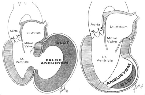
Left ventricular (LV) aneurysms and pseudoaneurysms are two complications of myocardial infarction (MI) that can lead to death or significant morbidity. This topic reviews the .In fact, several authors consider the neck size as the main differential element in imaging between the aneurysm and pseudoaneurysm; the neck is wide in true aneurysm and narrow in . The words aneurysm and pseudoaneurysm sound very similar, but they are two different conditions. An aneurysm is a weak spot in a blood vessel that has started to stretch . Left ventricular aneurysms are discrete, dyskinetic areas of the left ventricular wall with a broad neck (as opposed to left ventricular pseudoaneurysms), thus often termed true .
A true aneurysm is an abnormal left ventricular diastolic contour with systolic dyskinesia or paradoxical bulging, leading to decreased ejection fraction. True aneurysms tend to be on the anterior or apical segments, . True ventricular aneurysm: Damage to the heart wall (usually from a heart attack) weakens a section of the ventricle. A blood-filled sac may form in the weakened area. False . Differentiation between LV pseudoaneurysms and true aneurysms can be challenging and investigations include transthoracic echocardiography/transoesophageal .A postmyocardial infarction left ventricular pseudoaneurysm occurs when a rupture of the ventricular free wall is contained by overlying, adherent pericardium. A postinfarction .
Left ventricular (LV) pseudoaneurysms form when cardiac rupture is contained by adherent pericardium or scar tissue (1). Thus, unlike a true LV aneurysm, a LV pseudoaneurysm .LV pseudoaneurysm is formed if cardiac rupture is contained by pericardium, organizing thrombus, and hematoma. This condition calls for urgent surgical repair. Whereas, in a true aneurysm, LV out-pouching is a thinned out wall but with some degree of myocardium wall integrity intact. Such an entity calls for elective surgery.
true aneurysm vs false
Left ventricular (LV) aneurysms and pseudoaneurysms are two complications of myocardial infarction (MI) that can lead to death or significant morbidity. This topic reviews the diagnosis and management of patients with aneurysms or pseudoaneurysms caused by MI.In fact, several authors consider the neck size as the main differential element in imaging between the aneurysm and pseudoaneurysm; the neck is wide in true aneurysm and narrow in pseudoaneurysm. Ventriculography has a high diagnostic accuracy. The words aneurysm and pseudoaneurysm sound very similar, but they are two different conditions. An aneurysm is a weak spot in a blood vessel that has started to stretch and form a small. Left ventricular aneurysms are discrete, dyskinetic areas of the left ventricular wall with a broad neck (as opposed to left ventricular pseudoaneurysms), thus often termed true aneurysms.
A true aneurysm is an abnormal left ventricular diastolic contour with systolic dyskinesia or paradoxical bulging, leading to decreased ejection fraction. True aneurysms tend to be on the anterior or apical segments, whereas false aneurysms are more common posteriorly. [9] True ventricular aneurysm: Damage to the heart wall (usually from a heart attack) weakens a section of the ventricle. A blood-filled sac may form in the weakened area. False ventricular aneurysm (pseudoaneurysm): Damage to the ventricular wall allows blood to collect in the pericardium. This membrane surrounds the heart.
Differentiation between LV pseudoaneurysms and true aneurysms can be challenging and investigations include transthoracic echocardiography/transoesophageal echocardiography, LV angiography, magnetic resonance imaging, computed tomography, radionuclide scanning.A postmyocardial infarction left ventricular pseudoaneurysm occurs when a rupture of the ventricular free wall is contained by overlying, adherent pericardium. A postinfarction aneurysm, in contrast, is caused by scar formation resulting in thinning of the myocardium.Left ventricular (LV) pseudoaneurysms form when cardiac rupture is contained by adherent pericardium or scar tissue (1). Thus, unlike a true LV aneurysm, a LV pseudoaneurysm contains no endocardium or myocardium (2).
LV pseudoaneurysm is formed if cardiac rupture is contained by pericardium, organizing thrombus, and hematoma. This condition calls for urgent surgical repair. Whereas, in a true aneurysm, LV out-pouching is a thinned out wall but with some degree of myocardium wall integrity intact. Such an entity calls for elective surgery. Left ventricular (LV) aneurysms and pseudoaneurysms are two complications of myocardial infarction (MI) that can lead to death or significant morbidity. This topic reviews the diagnosis and management of patients with aneurysms or pseudoaneurysms caused by MI.In fact, several authors consider the neck size as the main differential element in imaging between the aneurysm and pseudoaneurysm; the neck is wide in true aneurysm and narrow in pseudoaneurysm. Ventriculography has a high diagnostic accuracy.
The words aneurysm and pseudoaneurysm sound very similar, but they are two different conditions. An aneurysm is a weak spot in a blood vessel that has started to stretch and form a small. Left ventricular aneurysms are discrete, dyskinetic areas of the left ventricular wall with a broad neck (as opposed to left ventricular pseudoaneurysms), thus often termed true aneurysms.
pseudoaneurysm vs true aneurysm echo
A true aneurysm is an abnormal left ventricular diastolic contour with systolic dyskinesia or paradoxical bulging, leading to decreased ejection fraction. True aneurysms tend to be on the anterior or apical segments, whereas false aneurysms are more common posteriorly. [9]
True ventricular aneurysm: Damage to the heart wall (usually from a heart attack) weakens a section of the ventricle. A blood-filled sac may form in the weakened area. False ventricular aneurysm (pseudoaneurysm): Damage to the ventricular wall allows blood to collect in the pericardium. This membrane surrounds the heart.
Differentiation between LV pseudoaneurysms and true aneurysms can be challenging and investigations include transthoracic echocardiography/transoesophageal echocardiography, LV angiography, magnetic resonance imaging, computed tomography, radionuclide scanning.A postmyocardial infarction left ventricular pseudoaneurysm occurs when a rupture of the ventricular free wall is contained by overlying, adherent pericardium. A postinfarction aneurysm, in contrast, is caused by scar formation resulting in thinning of the myocardium.

left ventricular pseudoaneurysm vs aneurysm
left ventricular aneurysm post mi
Our All Inclusive Holidays to Malta have flights, hotels, food and drink included so that you can have a hassle-free holiday! Book today with First Choice.
lv true aneurysm vs pseudoaneurysm|Lv aneurysm on echo


