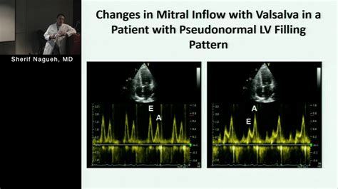
lv fill time | echocardiographic evaluation Lv filling lv fill time For normal cardiac performance, the left ventricle (LV) must be able to eject an adequate stroke volume at arterial pressure (systolic function) and . OMEGA's Seamaster Diver 300M collection includes a range of watches sized at 43.5 mm. All four models have much in common, including the use of black ceramic and the choice of movement: OMEGA’s Master Chronometer Calibre 8806.
0 · echocardiographic evaluation Lv filling
1 · Lv filling pressure measurement
2 · Lv filling pressure calculation
Approximately 70 hours. Bracelet. Oyster, three-piece solid links. Dial. Turquoise blue, Celebration motif. Certification. Superlative Chronometer (COSC + Rolex certification after casing) Turquoise blue dial. Named ‘Celebration’, this brand-new motif is available for the Oyster Perpetual 31, Oyster Perpetual 36 and Oyster Perpetual 41.
For normal cardiac performance, the left ventricle (LV) must be able to eject an adequate stroke volume at arterial pressure (systolic function) and .Values for average E/e′ ratio < 8 usually indicate normal LV filling pressures, values > 14 have high specificity for increased LV filling pressures. 1. E/e′ ratio is not accurate in normal . The gold standard for determining LV filling pressures, is performed invasively by measuring the left ventricular end-diastolic pressure (LVEDP) via cardiac cath. Another .

Recommendations for the Evaluation of Left Ventricular Diastolic Function by Echocardiography: An Update from the American Society of Echocardiography and the European Association of .
Diastolic dysfunction represents a combination of impaired left ventricular (LV) relaxation, restoration forces, myocyte lengthening load, and atrial function, culminating in increased LV filling pressures. Current Doppler .In patients with heart failure and reduced EF (HFrEF), the main goal is to estimate LV filling pressures and grade the degree of diastolic dysfunction (diastolic dysfunction is presumed to .
The decrease in rapid filling phase is demonstrated by a decrease E-wave velocity, usually < 50 cm/sec. The E-wave deceleration time (DT) will be elongated due to prolongation of impaired relaxation, usually >220 msec.
In the young up to the fourth decade of life, the left ventricle fills with a dominant early diastolic volume followed by a smaller late diastolic volume.Relative changes in chamber pressures can be studied using LV filling velocities, in different diastolic phases, obtained by spectral Doppler echocardiography, which has now become a well-established integral part of cardiac examination . For normal cardiac performance, the left ventricle (LV) must be able to eject an adequate stroke volume at arterial pressure (systolic function) and fill without requiring an elevated left atrial (LA) pressure (diastolic function). These systolic and diastolic functions must be adequate to meet the needs of the body both at rest and during stress.
Filling pressures: Normal; PW Doppler findings: E/A ratio < 1.0; Deceleration time > 240 msec; IVRT > 90 msec; Pulmonary Vein "a" wave flow reversal < 25 cm/sec; The stage I filling pattern represents impaired (slow) early left ventricular relaxation. At this point, the patient is usually asymptomatic, with normal filling pressures.Values for average E/e′ ratio < 8 usually indicate normal LV filling pressures, values > 14 have high specificity for increased LV filling pressures. 1. E/e′ ratio is not accurate in normal subjects, patients with heavy annular calcification, mitral valve and pericardial disease.The gold standard for determining LV filling pressures, is performed invasively by measuring the left ventricular end-diastolic pressure (LVEDP) via cardiac cath. Another method is by the pulmonary capillary wedge pressure (PCWP), which is an indirect measure of the mean LAP.
Recommendations for the Evaluation of Left Ventricular Diastolic Function by Echocardiography: An Update from the American Society of Echocardiography and the European Association of Cardiovascular Imaging.Diastolic dysfunction represents a combination of impaired left ventricular (LV) relaxation, restoration forces, myocyte lengthening load, and atrial function, culminating in increased LV filling pressures. Current Doppler echocardiography guidelines recommend using early to late diastolic transmitral flow velocity (E/A) to assess diastolic .In patients with heart failure and reduced EF (HFrEF), the main goal is to estimate LV filling pressures and grade the degree of diastolic dysfunction (diastolic dysfunction is presumed to be present in these patients) based on the parameters presented below and .
The decrease in rapid filling phase is demonstrated by a decrease E-wave velocity, usually < 50 cm/sec. The E-wave deceleration time (DT) will be elongated due to prolongation of impaired relaxation, usually >220 msec.In the young up to the fourth decade of life, the left ventricle fills with a dominant early diastolic volume followed by a smaller late diastolic volume.
Relative changes in chamber pressures can be studied using LV filling velocities, in different diastolic phases, obtained by spectral Doppler echocardiography, which has now become a well-established integral part of cardiac examination in health and disease. For normal cardiac performance, the left ventricle (LV) must be able to eject an adequate stroke volume at arterial pressure (systolic function) and fill without requiring an elevated left atrial (LA) pressure (diastolic function). These systolic and diastolic functions must be adequate to meet the needs of the body both at rest and during stress.Filling pressures: Normal; PW Doppler findings: E/A ratio < 1.0; Deceleration time > 240 msec; IVRT > 90 msec; Pulmonary Vein "a" wave flow reversal < 25 cm/sec; The stage I filling pattern represents impaired (slow) early left ventricular relaxation. At this point, the patient is usually asymptomatic, with normal filling pressures.Values for average E/e′ ratio < 8 usually indicate normal LV filling pressures, values > 14 have high specificity for increased LV filling pressures. 1. E/e′ ratio is not accurate in normal subjects, patients with heavy annular calcification, mitral valve and pericardial disease.
The gold standard for determining LV filling pressures, is performed invasively by measuring the left ventricular end-diastolic pressure (LVEDP) via cardiac cath. Another method is by the pulmonary capillary wedge pressure (PCWP), which is an indirect measure of the mean LAP.Recommendations for the Evaluation of Left Ventricular Diastolic Function by Echocardiography: An Update from the American Society of Echocardiography and the European Association of Cardiovascular Imaging.Diastolic dysfunction represents a combination of impaired left ventricular (LV) relaxation, restoration forces, myocyte lengthening load, and atrial function, culminating in increased LV filling pressures. Current Doppler echocardiography guidelines recommend using early to late diastolic transmitral flow velocity (E/A) to assess diastolic .
In patients with heart failure and reduced EF (HFrEF), the main goal is to estimate LV filling pressures and grade the degree of diastolic dysfunction (diastolic dysfunction is presumed to be present in these patients) based on the parameters presented below and .The decrease in rapid filling phase is demonstrated by a decrease E-wave velocity, usually < 50 cm/sec. The E-wave deceleration time (DT) will be elongated due to prolongation of impaired relaxation, usually >220 msec.In the young up to the fourth decade of life, the left ventricle fills with a dominant early diastolic volume followed by a smaller late diastolic volume.
echocardiographic evaluation Lv filling
Lv filling pressure measurement
Lv filling pressure calculation
Dior 30 Montaigne Card Holder. Save Search. Recent Sales. Dior Nude Leather 30 Montaigne 5 Pocket Card Holder. Get Updated with New Arrivals. Save "Dior 30 Montaigne Card Holder", and we’ll notify you when there are new listings in this category. Save Search. Christian Dior for sale on 1stDibs.
lv fill time|echocardiographic evaluation Lv filling


